What are the types of microscopes?
Microscopes are equipment used to magnify small objects to help you see some details that you can see with your naked eyes.
In microbiology, telescopes are used in different applications among them the study of micro-organisms that are too tiny to be visible to the human eye.
There are several types of microscopes suitable for different applications. Typically, microscopes run on batteries or are powered by mechanical mechanisms to magnify the specimen.
In an event where the specimen is handled incorrectly on inappropriately, the microscope can distort the image and give false results. So, handling the specimen with caution and using the right microscope is important.
Types of Microscopes Description and Uses
Here are 5 different microscope types with their specific features and uses.
Simple Microscope

A simple microscope comes with large magnifying optics and a shorter focal length that features a small convex mirror with a miniature focal area. The most common simple microscopes are handheld lenses and eyepiece lenses.
When you put a specimen close to the lens of a simple microscope, a focus is created then the original object gets magnified and more erect. The microscope then focuses on a particular portion of the object by bringing two edges of the lens together. This creates a tiny but more focused image of the object than viewing the whole object.
Simple microscopes have one magnification level only depending on the type of lens used. This makes them ideal for reading and magnifying less complex items. For example, you can use the magnifying lens to zoom in on details when reading a map.
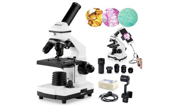
Compound Light Microscope
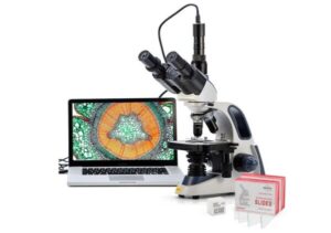
A compound microscope is the most popular type of microscope you’ll find being used by most people. This type of microscope has a lens on it that features a compound medium in between. The compound medium allows for smooth magnifications on a very clear scale.
While simple microscopes use natural light to magnify an object, compound microscopes require illumination to view the specimen. Here are the basic specifications found in a compound microscope.
- Magnification makes the specimen look larger when you look through the microscope’s lenses. The magnification strength ranges from 40x to 400x depending on the specific model.
- Resolution refers to how goof and clear the image is captured by the microscope’s lens. A compound microscope with a higher resolution will provide clearer and more detailed images. Additionally, it has greater visual clarity since it has additional layers of magnification.
- You can stain the specimen to achieve excellent contrast so the colors in the specimen will stand out when you view it in the microscope.
A compound microscope is extremely helpful for research purposes. Most of these microscopes are used when viewing scientific specimens for educational or research purposes. They are mainly used in laboratories and medical schools.
Stereo Microscope

A stereo/stereoscopic microscope is specially designed for low magnification of biological specimens. The microscope works by reflecting light off the specimen’s surface instead of transmitting light through its medium.
In most cases, stereo microscopes are used in chemistry laboratories where detailed, 3D images are needed. This type of microscope produces high-quality images that can only be found in electron microscopes or other high-powered microscopes.
Stereo microscopes are a favorite among most people because of the quality of images they produce. Additionally, these microscopes are relatively affordable and require minimal maintenance which makes them economical to run. You can use them to view manufacturing materials, circuit board work, inspection, and dissection.
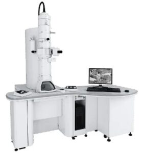
Scanning Electron Microscope (SEM)

A scanning electron microscope is designed to produce images of a specimen by scanning it with a high-strength beam of electrons. The electrons will interact with the atoms within the specimen and create different signals which carry the data concerning the structure and topography of the specimen in observation. This microscope produces highly accurate images. You can also view the images in high resolution through the microscope eyepiece or magnifier.
To obtain accurate results from this microscope, the specimen should have electrical conduction. This creates a surface for electrons to bounce off and create a clear image. You can coat the specimen with a thin layer of metal such as gold to make it electrically conductive.
You can also use a number of techniques to enhance the image quality. This includes fluorescence imaging, multi-beam scanning, and tip electron microscopy among others. Furthermore, you should always make sure that the microscope is in the best working condition. This improves the quality of the images that you get.
Scanning electron microscopes are used for art, medicine, material science, forensic investigation, soil, and rock sampling, and semi-conductor inspection as well.
Transmission Electron Microscope (TEM)
With transmission electron microscopes, the microscope uses a beam of electrons that are transmitted through an unstained specimen to generate an optical image. Unlike SEMs microscopes where electrons bounce off when they hit the specimen, the electrons in TEMs pass through the specimen. Typically, the specimen is ultra-thin and less than 50micrometers thick.
What makes TEM microscopes stand out among the rest is the high magnification power. They have an amazing optical power that’s possibly 10,000x better than that in optical microscopes. This is particularly important for researchers looking for every detail in small specimens.
However, these types of microscopes have their drawbacks. They are very expensive because of the sophisticated features they have. They are accessible to scientists only, but not students. Plus, samples to be viewed require detailed preparation before they can be placed on the microscope and you can’t use the microscope to observe a living sample such as protozoa.
TEM microscopes are mainly used in the field of nanotechnology, life science, biological and material research, gemology, metallurgy and medical research.
Final Thoughts
Microscopes are highly beneficial in the field of science. They are not only essential for medical students but also for scientists. With technological advancements being the order of the day, it might just take us a few years before microscopes are transformed into new and much better equipment with greater potential than what is present today.
See Also